Every dentist who has delivered a nasopalatine nerve block knows why patients dread the anterior palate injection. The tissue is dense, tightly bound to bone, and richly innervated, which turns even a careful stick into one of the more painful moments in dental care. Understanding the pathophysiology behind that response is the first step toward making the injection more tolerable for the patient in the chair.
Cryo-anesthesia offers a targeted way to blunt the sharp moment without changing the underlying anesthetic protocol. Rapid surface cooling slows nerve conduction in the mucosa before the needle enters, which reduces the intensity of the initial signal. The result is a smoother anterior injection that lands closer to what patients actually expect from modern dental care.
What Role Does the Nasopalatine Nerve Play in Anterior Palatal Pain?
The nasopalatine nerve carries sensory information from the anterior palatal mucosa behind the maxillary central incisors, which makes it central to any pain response in that region. Dense innervation and tightly attached mucosa combine to make the nasopalatine block one of the most sensitive injection sites in the mouth. Surface cooling can numb the pain signal by slowing conduction in the fibers before the needle enters the tissue.
Anatomy and Innervation of the Nasopalatine Nerve
The nasopalatine nerve begins as a branch of the maxillary division of the trigeminal nerve inside the pterygopalatine fossa. It descends through the nasal cavity, runs along the nasal septum, and exits through the incisive foramen behind the maxillary central incisors. From there it supplies sensory innervation to the anterior third of the hard palate mucosa.
Two nasopalatine nerves, one from each side, converge at the incisive foramen and often join into a single trunk. The convergence explains why a single injection at the incisive papilla can anesthetize the anterior palatal mucosa bilaterally in most patients. Anatomic variation exists, however, and some patients require slight repositioning to achieve full coverage.

The fibers themselves are primarily sensory, carrying pain, temperature, and touch information from the anterior palate. Small autonomic components also travel with the nerve, contributing to vascular and mucous gland control in the region. The mix of sensory density and small autonomic input shapes how the tissue responds to any stimulus.
Furthermore, the mucosa overlying the incisive foramen is tightly bound to the underlying bone with minimal submucosal space. The dense attachment limits how much anesthetic solution can spread without generating significant tissue pressure. Anatomy alone explains a large portion of why the injection feels the way it does at the chair.
Why the Anterior Palate Delivers Such Sharp Pain During Injection
The anterior palatal mucosa is one of the most densely innervated regions in the entire oral cavity. Nerve fibers sit close to the surface, and the tissue itself has little room to accommodate the volume of an incoming anesthetic solution. Each contribution stacks to make the injection more intense than a comparable stick elsewhere in the mouth.
Pressure at the site is the second major driver of the sharp response patients report. Fluid injected into tightly bound tissue has nowhere to go, so the anesthetic pushes back against the mucosa itself.
Speed of injection also shapes how the moment feels for the patient in the chair. Rapid delivery increases tissue distension and amplifies the mechanical pain component of the stick. Slower, controlled delivery reduces that component, though it does not eliminate the initial needle contact.
Meanwhile, patient anxiety compounds every physical factor listed above during a real appointment. Anticipation of a painful stick raises baseline pain scores before the needle even touches the tissue. Reducing the sharp initial moment therefore matters both physically and psychologically for the anterior palatal injection.
How Cryo-Anesthesia Modulates Nasopalatine Nerve Signaling
Cryo-anesthesia modulates nasopalatine nerve signaling by lowering surface temperature at the injection site within seconds of application. With the right level of cooling, those signals can be temporarily stopped altogether. The effect covers the exact moment when the needle would otherwise trigger the sharpest response from the patient.
Rapid evaporative cooling reaches the superficial sensory fibers first, which are the same fibers responsible for the initial pain burst. Tissue depth influences how far cooling penetrates, with thinner palatal tissues allowing vapocoolants to cool more deeply throughout the treatment area. The layered effect means that cryo-anesthesia functions as a fast-acting topical anesthesia, preparing the site for a more comfortable local anesthetic injection.
The cooling window fades within seconds, matching the physiologic moment of the stick almost exactly. Longer procedures still rely on the injected anesthetic for sustained numbing, which is the intended clinical division of labor. Surface cooling handles the initial mucosal contact, and the local agent handles everything after for the duration of the visit.
However, technique matters more than product choice when applying cooling to the anterior palate at chairside. Timing, distance, and duration all shape whether the physiologic effect lands where it needs to.
Applying Surface Cooling to the Anterior Palatal Injection
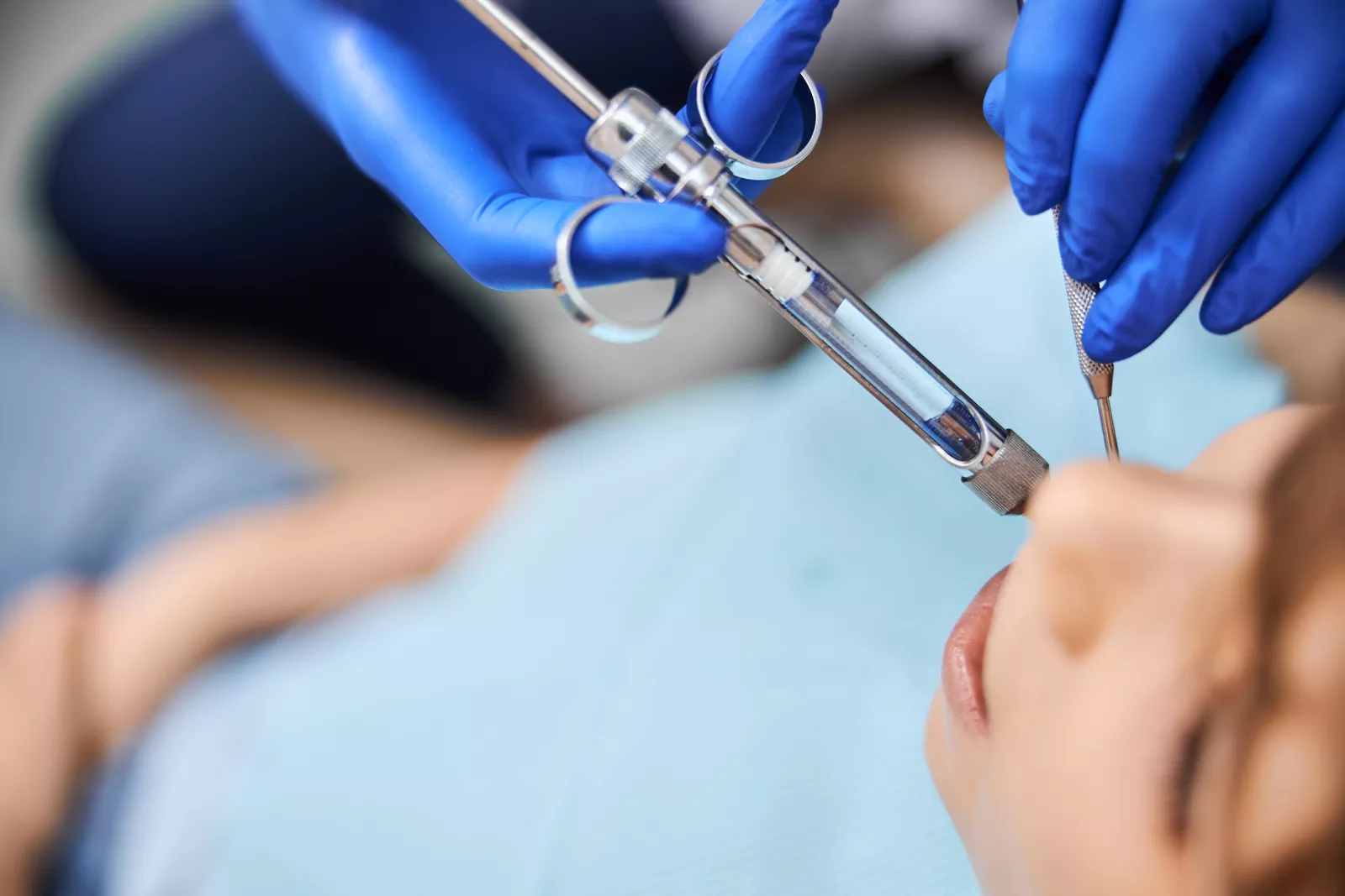
Surface cooling for the anterior palatal injection works best when applied directly over the incisive papilla just before the stick. The nasopalatine nerve exits at that exact spot, so cooling the papilla targets the fibers most responsible for the sharp response. Positioning the spray perpendicular to the mucosa gives the most consistent coverage at the papilla.
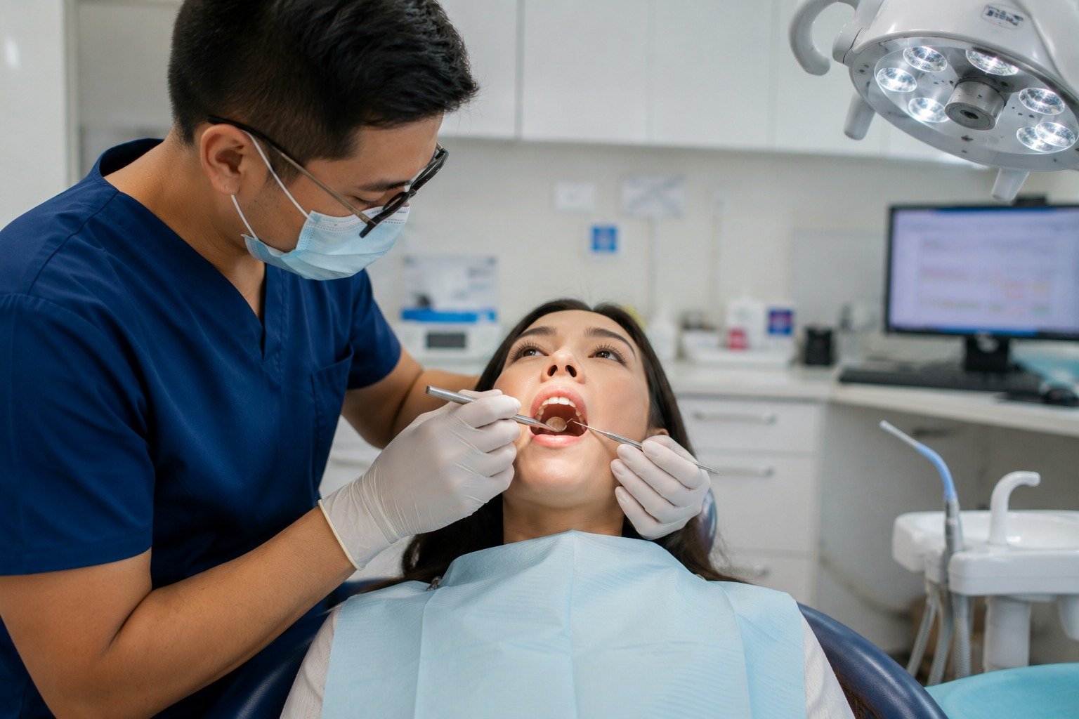
Application duration is typically around one second for the anterior palate. Longer application can cause frostbite risk in the dense, tightly bound mucosa, which has less blood flow than other oral sites.
The injection should follow immediately after the cooling application ends at the site. Waiting even 5seconds allows the surface temperature to rebound and reduces the effect on the initial stick. Clinical protocols that pair the two steps into a single continuous motion produce the smoothest anterior palatal injections.
In addition, patient positioning matters more at the anterior palate than at most other sites in the mouth. A slight head tilt gives clearer access to the papilla for both the spray and the needle. Assistants who own the cooling step tend to get more consistent results than dentists attempting to administer both the cooling and local anesthetic injection themselves.
Ready to Take the Sharp Edge Off Anterior Palatal Injections?
Anterior palatal injections are among the most difficult moments in dental care for patients and clinicians alike. Cryo-anesthesia gives your team a fast, targeted way to blunt the initial stick without changing the anesthetic protocol. Small workflow adjustments deliver noticeably calmer visits from the first application forward.
DentalJect targets the incisive papilla in seconds, cooling the mucosa right before the nasopalatine block enters the tissue. The device pairs cleanly with your existing anesthetic sequence and drops in without any protocol rewrite. Reach out to the VCS Medical team about adding DentalJect to your anterior palatal injection workflow.
Ready to start the No-Wait Injection Protocol?
Add DentalJect to your practice with the Intro Bundle.
Get the Intro Bundle